GE 3T Keck Protocol Development
High Contrast & Quantitative UTE Imaging in the Spine
MRI Protocol Objectives
High contrast UTE imaging of the cartilaginous endplate (CEP) and cortical bone.
Quantitative UTE imaging of the spine using UTE-T2, UTE-MT, UTE-tricomponent and UTE-multicompartment imaging.
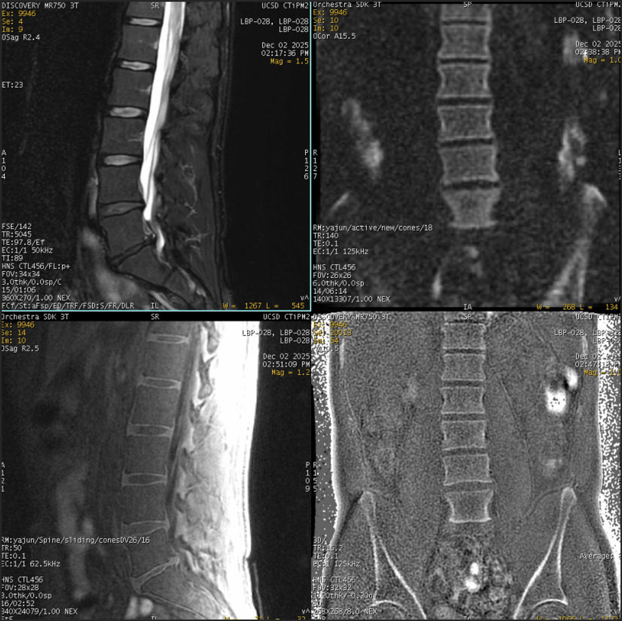
Example MR Images
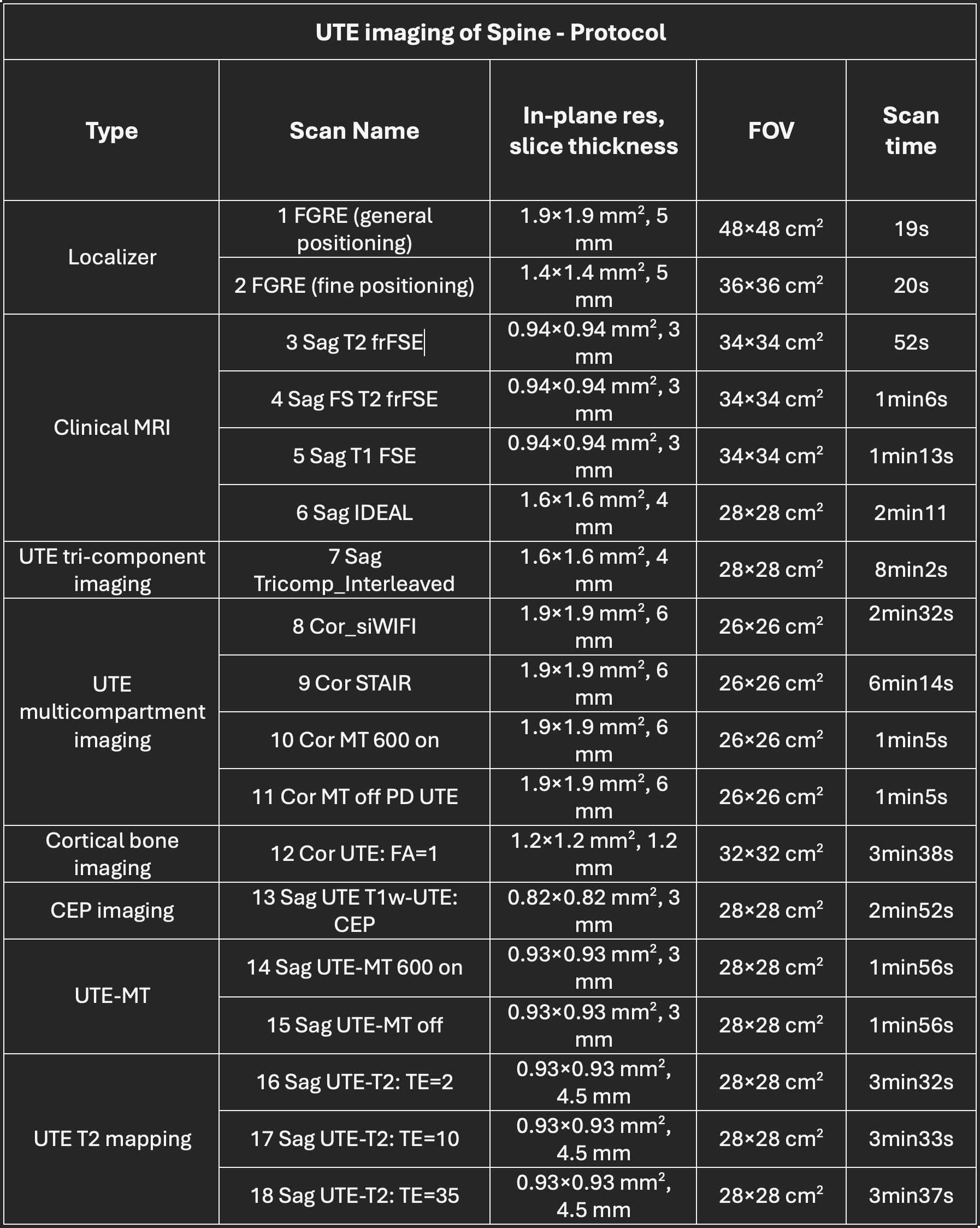
Protocol Parameter Overview
Protocol Steps
Set up the spine coil. Imaging with feet first and using coil elements 4-6 for all the scans.
The imaging planes of all the sequences are labeled in the names, except for the localizers.
Patient Positioning
Position the patient in the feet-first, supine position on the scanner bed.
Align the top of the anterior superior iliac spine with the central region of coil elements 4 and 5.
Place a bolster pad under the knees.
Physiological Gating
Not applicable
Post-Processing Support
Dr. Yajun Ma and his research team can provide customized MATLAB code for data processing.
Contact: Yajun Ma - yam013@health.ucsd.edu
Acknowledgement
Thanks to Dr. Yajun Ma and his team for their effort in developing this exciting protocol on the GE MR750 3T Keck MRI scanner at the UCSD CFMRI.
UTE imaging of Hemophilic Arthropathy
MRI Protocol Objectives
UTE quantitative susceptibility mapping (QSM) of ankle joints with Hemophilic Arthropathy (HA)
UTE QSM of knee joints with HA
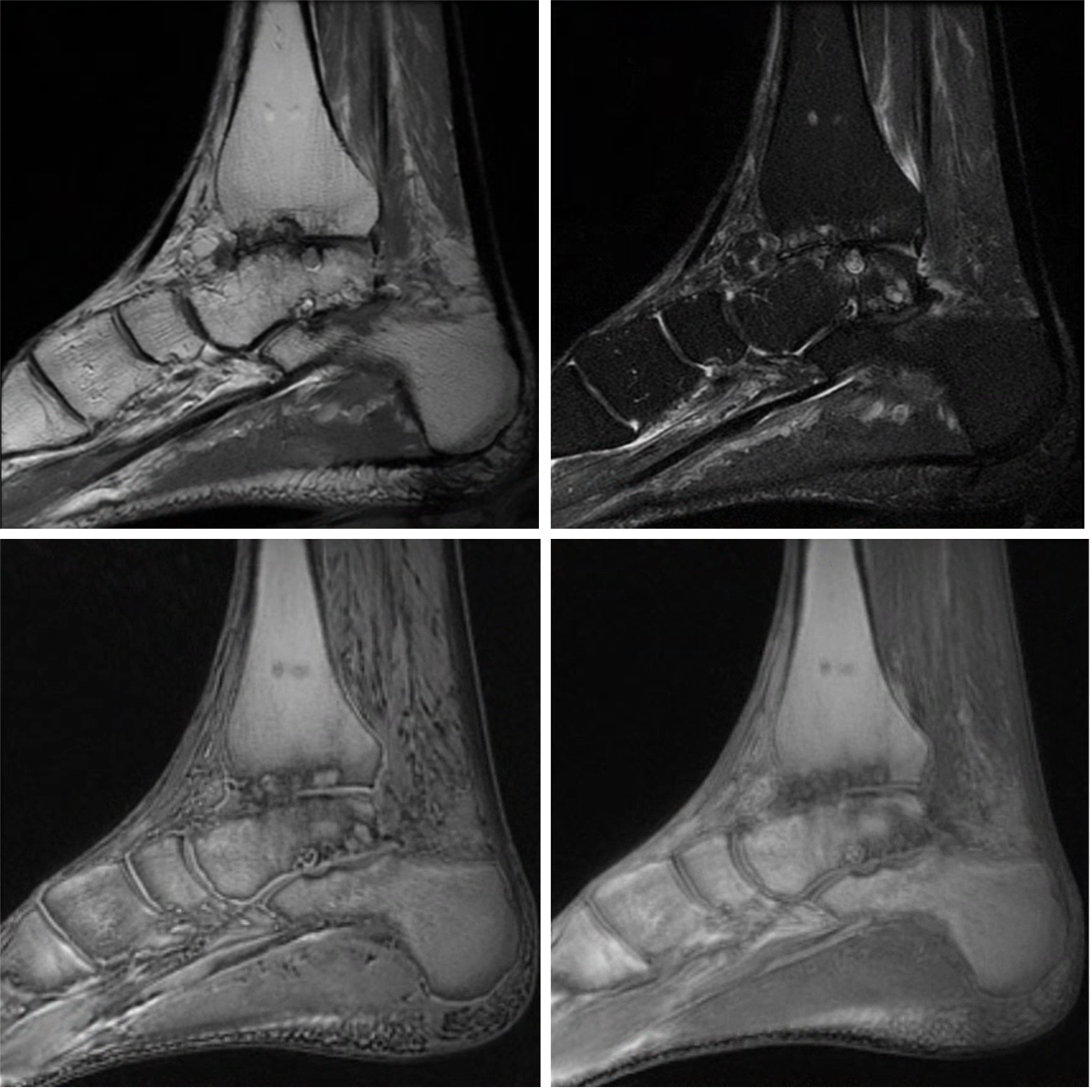
Example MR Images
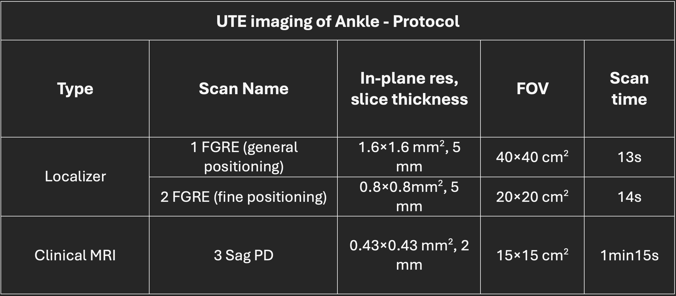
Protocol Parameter Overview
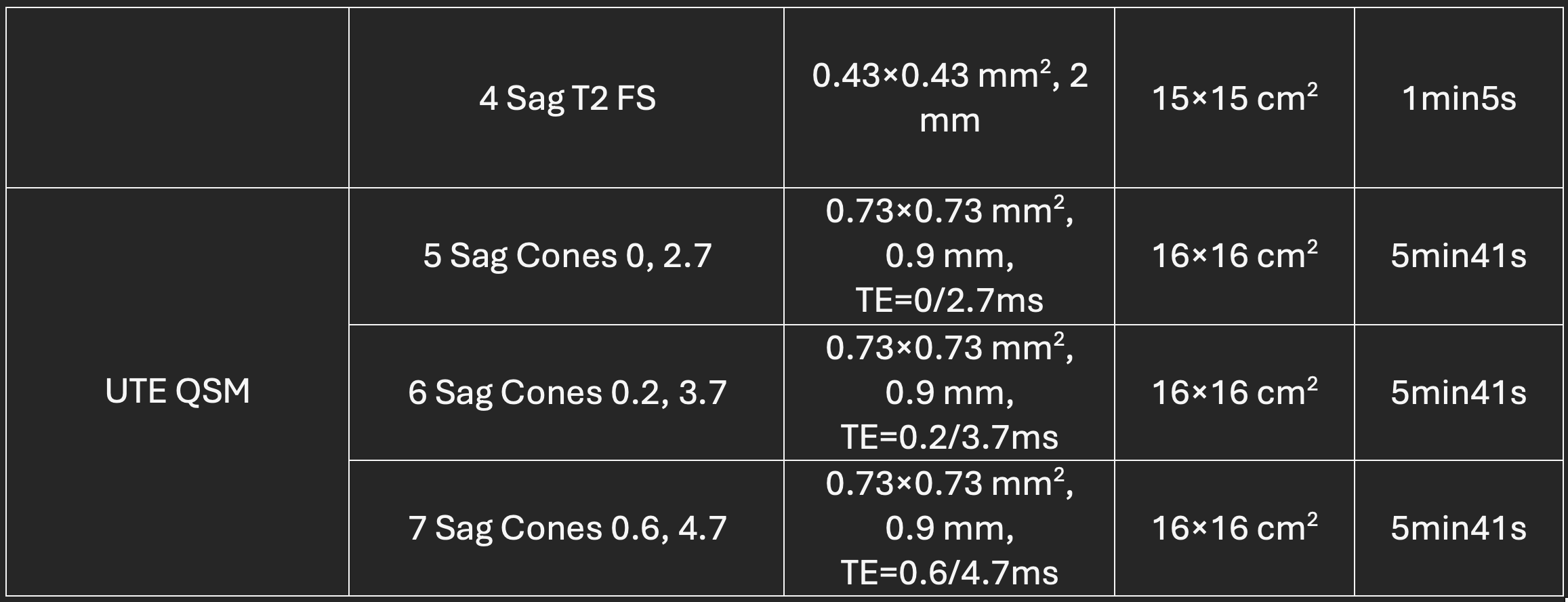
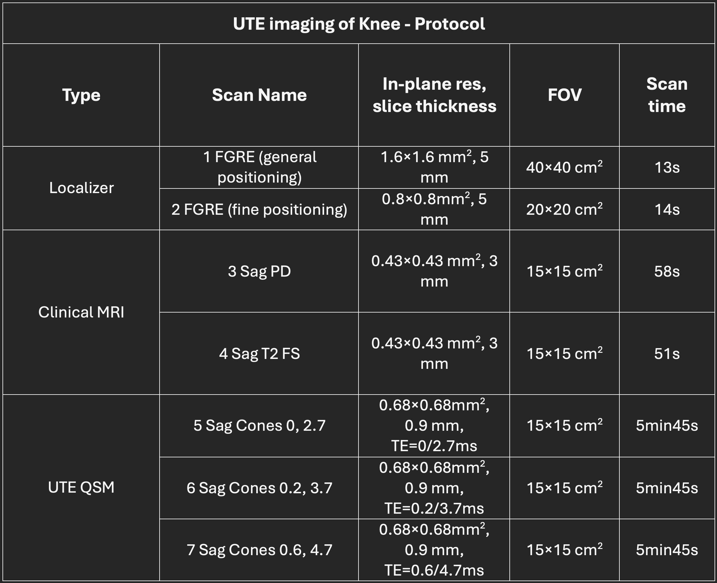
Protocol Steps
Ankle Imaging
Set up the ankle coil. Imaging should be performed with the patient positioned feet first.
The imaging planes of all the sequences are labeled in the names, except for the localizers.
For QSM imaging, the first sequence should be scanned in auto mode. The second and third sequences should be scanned using manual prescan to ensure that all calibration parameters (e.g., R1, R2, and center frequency) remain the same as in the first scan.
Knee Imaging
Set up the knee coil. Imaging should be performed with the patient positioned feet first, with the inferior border (apex) of the patella aligned with the center marker of the knee coil.
The imaging planes of all the sequences are labeled in the names, except for the localizers.
For QSM imaging, the first sequence should be scanned in auto mode. The second and third sequences should be scanned using manual prescan to ensure that all calibration parameters (e.g., R1, R2, and center frequency) remain the same as in the first scan.
Positioning
Feet first
Inferior border (apex) of the patella aligned with the center marker of the knee coil for knee imaging.
Place pads under and above the knee and ankle to keep them secure and stable during the scan.
Physiological Gating
Not Applicable
Post-Processing Support
Dr. Yajun Ma and his research team can provide customized MATLAB code for data processing.
Contact: Yajun Ma - yam013@health.ucsd.edu
Acknowledgement
Thanks to Dr. Yajun Ma and his team for their effort in developing this exciting protocol on the GE MR750 3T Keck MRI scanner at the UCSD CFMRI.